Microdisplay can monitor brain activity in real-time
American team develops LED-based device to visualise brain activity to guide neurosurgeons during brain surgery
Engineers and physicians from University of California San Diego and Massachusetts General Hospital (MGH) have developed a thin film display device that combines an electrode grid and special GaN LEDs that can both track and produce a visual representation of the brain’s activity in real-time during surgery.
The device is designed to help neurosurgeons monitor brain states during surgical interventions to remove brain lesions including tumors and epileptic tissue.
The team, led by Shadi Dayeh, a professor in the department of Electrical and Computer Engineering at UC San Diego, describes the work in the April 24 issue of the journal Science Translational Medicine.
Each LED in the device mirrors the activity of a few thousand neurons. The researchers have shown that the device can track and display neural activity in the brain corresponding to different areas of the body. In this case, the LEDs developed by the team light up red in the areas that need to be removed by the surgeon. Surrounding areas that control critical functions and should be avoided show up in green.
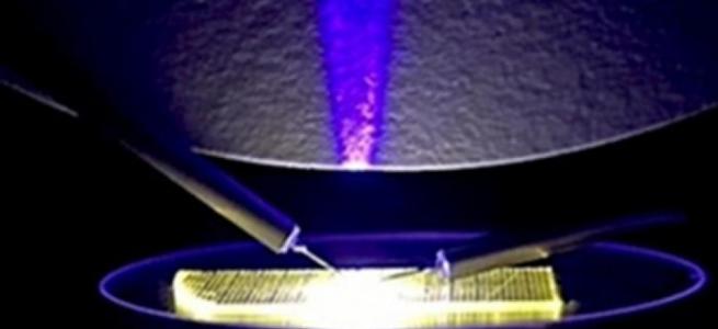
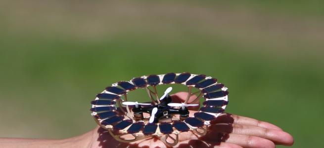
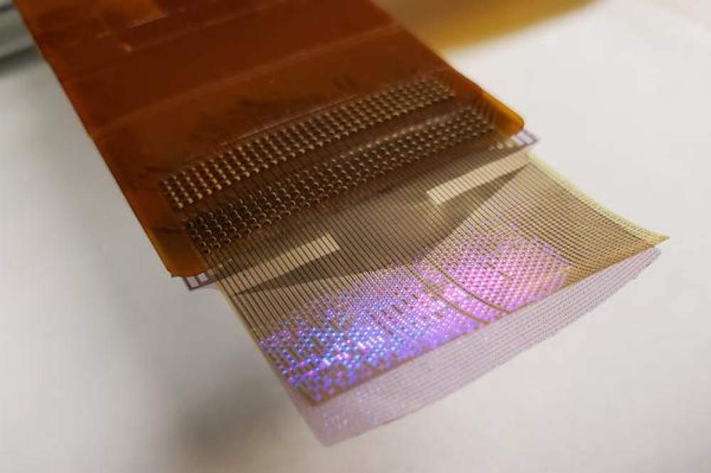
Dayeh and his team used their expertise in working with GaN to develop a manufacturing technique for high-efficiency LEDs that do not heat up when they light up and do not damage brain tissues. The material itself is grown on a flat and rigid substrate (Qromis substrate technology). Dayeh’s team was able to embed thousands of LEDs in flexible films and release them from the substrate in the form of a flexible display panel. Researchers then used inkjet printing to deposit quantum dot inks on the surface of the LEDs to convert their blue light to multiple other colors. “This enables richer and more nuanced visual representation of neural activity patterns,” said Dayeh.
“These GaN-based inorganic micro-LEDs, substantially brighter and power-efficient than organic LEDs, can maintain clear visibility under surgical lights that may exceed the brightness of direct sunlight. The iEEG microdisplay, just a few tens of microns thick, captures brain activity at 20,000 samples per second across thousands of channels and visualizes it at a video rate of 40 Hz. This enables precise and real-time displays of cortical dynamics during critical surgical interventions,” said Youngbin Tchoe, co-inventor, formerly a postdoc in the Dayeh group at UC San Diego and now an assistant professor at Ulsan National Institute of Science and Technology.
Then researchers installed the LEDs on top of another innovation from the Dayeh lab, the platinum nanorod electrode grid (PtNRGrid). Using the PtNRGrids since 2019, Dayeh’s team pioneered human brain and spinal cord mapping with thousands of channels to monitor brain neural activity. The PtNRGrid also includes perforations, which enable physicians to insert probes to stimulate the brain with electrical signals, both for mapping and for therapy.
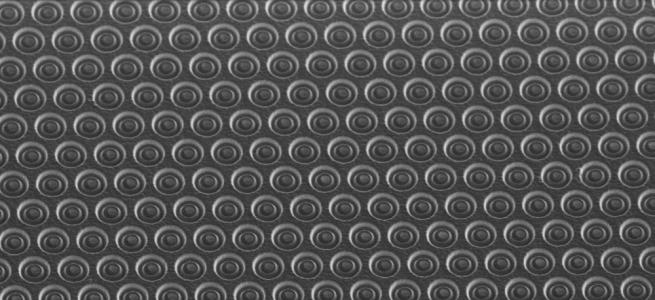
The microdisplays measure 5 by 5 square millimeters and 32 by 32 square millimeters and include either 1024 or 2048 of these LEDs, laminated on the back of the PtNRGrid. In addition to the LEDs, the device includes acquisition and control electronics as well as software drivers to analyze and project cortical activity directly from the surface of the brain.
Dayeh’s team is now working to build a microdisplay that will include 100,000 LEDs, with a resolution equivalent to that of a smartphone screen. Each LED in those displays would reflect the activity of a few hundred neurons. These brain microdisplays will cost a fraction of a high-end smartphone.
This brain microdisplay will also include a foldable portion. This would allow surgeons to operate within the foldable portion and monitor the impact of the procedure as the other, unfolded portion of the microdisplay shows the status of the brain in real time.
Researchers are also working on one limitation of the study. The close proximity of the LED sensors and the PtNRGrids led to a slight interference and noise in the data. The team plans to build customised hardware to change the frequency of the pulses that turn on the LEDs to make it easier to screen out that signal, which is not relevant to the brain’s electrical activity.
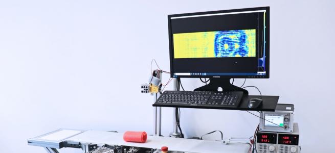
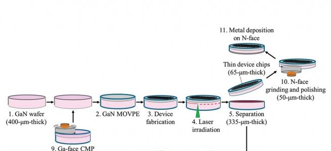
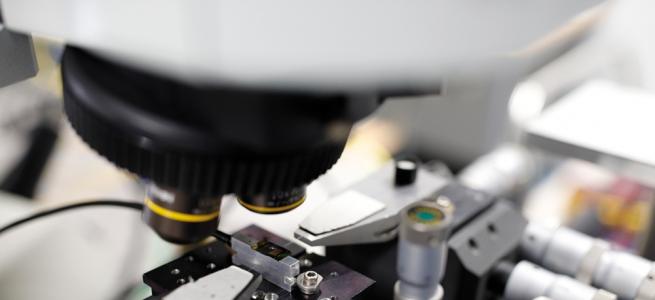
The brain microdisplay was fabricated in the Nano3 cleanroom facilities at UC San Diego’s Qualcomm Institute.